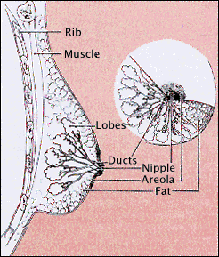
It is important for women to become familiar with the normal anatomy and physiology (function) of their breasts so that they can recognize early signs of possible abnormalities. This section outlines basic information on breast composition, development, and typical changes from puberty to pregnancy to menopause.
The breast is a mass of glandular, fatty, and fibrous tissues positioned over the pectoral muscles of the chest wall and attached to the chest wall by fibrous strands called Cooper’s ligaments. A layer of fatty tissue surrounds the breast glands and extends throughout the breast. The fatty tissue gives the breast a soft consistency.

Image courtesy of NCI/NIH
The glandular tissues of the breast house the lobules (milk producing glands at the ends of the lobes) and the ducts (milk passages). Toward the nipple, each duct widens to form a sac (ampulla). During lactation, the bulbs on the ends of the lobules produce milk. Once milk is produced, it is transferred through the ducts to the nipple.
The breast is composed of:
- milk glands (lobules) that produce milk
- ducts that transport milk from the milk glands (lobules) to the nipple
- nipple
- areola (pink or brown pigmented region surrounding the nipple)
- connective (fibrous) tissue that surrounds the lobules and ducts
- fat
Visit Healthline.com's free interactive Human Female Chest in 3D.
Arteries carry oxygen rich blood from the heart to the chest wall and the breasts and veins take de-oxygenated blood back to the heart. The axillary artery extends from the armpit and supplies the outer half of the breast with blood; the internal mammary artery extends down from neck and supplies the inner portion of the breast.
Human breast tissue begins to develop in the sixth week of fetal life. Breast tissue initially develops along the lines of the armpits and extends to the groin (this is called the milk ridge). By the ninth week of fetal life, it regresses (goes back) to the chest area, leaving two breast buds on the upper half of the chest. In females, columns of cells grow inward from each breast bud, becoming separate sweat glands with ducts leading to the nipple. Both male and female infants have very small breasts and actually experience some nipple discharge during the first few days after birth.
Female breasts do not begin growing until puberty—the period in life when the body undergoes a variety of changes to prepare for reproduction. Puberty usually begins for women around age 10 or 11. After pubic hair begins to grow, the breasts will begin responding to hormonal changes in the body. Specifically, the production of two hormones, estrogen and progesterone, signal the development of the glandular breast tissue. This initial growth of the breast may be somewhat painful for some girls. During this time, fat and fibrous breast tissue becomes more elastic. The breast ducts begin to grow and this growth continues until menstruation begins (typically one to two years after breast development has begun). Menstruation prepares the breasts and ovaries for potential pregnancy.
| Before puberty | Early puberty | Late puberty |
| the breast is flat except for the nipple that sticks out from the chest | the areola becomes a prominent bud; breasts begin to fill out | glandular tissue and fat increase in the breast, and areola becomes flat |


