


The following case shows magnetic resonance (MR) breast imaging of a patient with a benign fibroadenoma (a) and invasive lobular carcinoma (b). The two images (left and right) are taken from different planes before contrast injection (gadolinum) and then after contrast injection. The final set of pictures shows the post-contrast images subtracted from the pre-contrast images in order to display regions that are highlighted by the contrast. In particular, the subtracted images highlight the spiculated area that shows invasive lobular carcinoma. A subsequent biopsy on the patient confirmed the presence of cancer.
Before contrast injection:
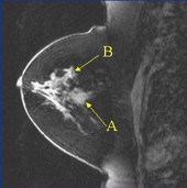
A: Fibroadenoma
B: Invasive Lobular Carcinoma
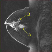
Five minutes after contrast injection:
A: Fibroadenoma
B: Invasive Lobular Carcinoma
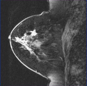
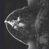
Subtracted images (only the cancer is visible):
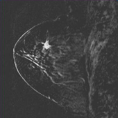
A: Fibroadenoma
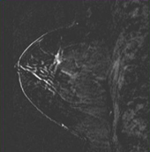
B: Invasive Lobular Carcinoma
Images are courtesy of Robert A. Pooley, PhD and John Knudsen, MD of the Mayo Clinic Jacksonville, David Thomasson, PhD of Georgetown University Hospital and Siemens Medical Solutions.
