


Over the years, medical imaging has become a vital part in the early detection, diagnosis and treatment of cancer. In some cases medical imaging is the first step in preventing the spread of cancer through early detection and in many cases makes it possible to cure or eliminate the cancer altogether. Computed Tomography (CT) imaging, Magnetic Resonance (MR) imaging, Mammography, Nuclear Medicine (NM) imaging and Ultrasound (US) imaging and X-ray imaging are all very important tools in the fight against cancer.
After cancer has been diagnosed, imaging is often used to follow the course of cancer treatment and to monitor the growth or remission (disappearance of the signs of cancer). Medical imaging is being used more frequently to build precise computer models which allow doctors to guide exact radiation treatment of cancer (click here to go to the section on radiation treatment planning using CT and MR data).
CT and MR are both exceptionally good at imaging soft tissue structures, and thus are excellent for detecting and diagnosing tumors and to differentiate if they are benign or malignant.
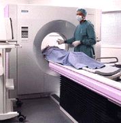
CT imaging is typically the method of choice for imaging tumors of the lungs, abdomen, liver, kidneys, pancreas and pelvis. This is due to its fast speed of data acquisition and thus its ability to minimize disturbances (artifacts) in an image which may be caused by patient motion, breathing or peristalsis (involuntary wavelike contraction of gastro-intestinal organs). CT can also be used to look for tumors in the brain and spine. CT scanning is also being used more and more to guide biopsy (sampling) of tumors in order to test if the tumor is benign or malignant. Newer spiral CT scanners are being equipped with so called "Interventional CT" packages to allow real time CT imaging for the guidance of biopsy.
 |
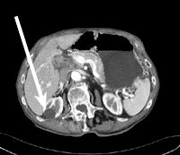 | |
| The radiologist uses "real-time CT guidance" to guide biopsy of a tumor,he can watch on the progress of the needle placement on the monitor as he approaches the lung tumor. | Axial CT image of the liver and kidneys shows a benign (non-cancerous) cyst (arrow) |
MR imaging has excellent contrast resolution, meaning it can show a subtle tumor or soft-tissue growth with exceptional clarity. Thus, MR is often the method of choice for diagnosing brain tumors and for searching for metastases (cancers which have spread from another organ) of the spine. MR is also an excellent method for imaging tumors in joints and bones. New, faster MR acquisition techniques are also allowing MR to be used more often to image cancer of the liver and abdomen.
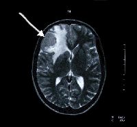 |
This axial MR image of the brain shows a large tumor in upper left corner of image (arrow) |
Nuclear Medicine imaging (including Positron Emission Tomography, PET) is often used to search for cancer in the organs and the skeletal system. The nuclear medicine "bone scan" is a routine examination for this purpose.
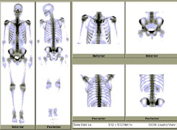 |
| This nuclear medicine bone scan is normal and shows no cancer |
Ultrasound imaging is a versatile technique for searching for abdominal tissue masses if a patient has symptoms of abdominal pain. Ultrasound can also be used to guide biopsy (sampling) of tumors to test if they are benign or malignant.
 |
| This digital x-ray image shows a normal stomach |
Various types of x-ray imaging are central to diagnosis of many types of cancer. Fluoroscopy is useful for detecting disorders and tumors in the upper gastro-intestinal (GI) system (for example, the stomach and intestines). Chest x-ray is often the first step in detecting lung cancer and mammography is the primary means for detecting and diagnosing breast cancer.
Updated: June 9, 2008
