


Unlike other medical imaging techniques, such as conventional x-ray imaging (radiography), CT enables direct imaging and differentiation of soft tissue structures, such as liver, lung tissue, and fat. CT is especially useful in searching for large space occupying lesions, tumors and metastasis and can not only reveal their presence, but also the size, spatial location and extent of a tumor.
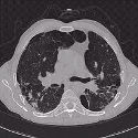
High resolution axial CT image of the chest showing the vessels of the heart (center of image) and pneumonia in the both lungs.
CT imaging of the head and brain can detect tumors, show blood clots and blood vessel defects, show enlarged ventricles (caused by a build up of cerebrospinal fluid) and image other abnormalities such as those of the nerves or muscles of the eye.
Due to the short scan times of 500 milliseconds to a few seconds, CT can be used for all anatomic regions, including those susceptible to patient motion and breathing. For example, in the thorax CT can be used for visualization of nodular structures, infiltrations of fluid, fibrosis (for example from asbestos fibers), and effusions (filling of an air space with fluid).
CT has been the basis for interventional work like CT guided biopsy and minimally invasive therapy. CT images are also used as basis for planning radiotherapy cancer treatment. CT is also often used to follow the course of cancer treatment to determine how the tumor is responding to treatment.
CT imaging provides both good soft tissue resolution (contrast) as well as high spatial resolution. This enables the use of CT in orthopedic medicine and imaging of bony structures including prolapses (protrusion) of vertebral discs, imaging of complex joints like the shoulder or hip as a functional unit and fractures, especially those affecting the spine. The image postprocessing capabilities of CT - like multiplanar reconstructions and 3-dimensional display (3D) - further enhance the value of CT imaging for surgeons. For instance, 3-D CT is an invaluable tool for surgical reconstruction following facial trauma.
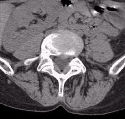
Axial CT image of the lumbar spine showing a slight prolapse of the disk impinging on the spinal cord.
CT is becoming the method of choice for imaging trauma patients. CT exams are fast and simple and enable a quick overview of possibly life-threatening pathology and rapidly enables a dedicated surgical treatment.
With the advent of spiral CT, the continuous acquisition of complete CT volumes can be used for the diagnosis of blood vessels with CT Angiography. For instance, abdominal aortic aneurysms, the renal arteries, the carotids vessels and the Circle of Willis can all now be quickly imaged with CT with minimal intervention.
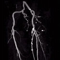
CT angiography image of the femoral arteries showing multiple calcifications (bright white lumps) on the femoral branch on the right
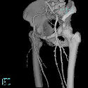
3D Surface reconstruction of the same femoral arteries showing their position relative to the pelvis and the femurs
Due to the short total acquisition time of spiral CT, imaging of the liver is now possible in different contrast enhancement phases. These so-called "multi-phase" studies offer a step towards differential diagnosis of lesions in the liver. In other words, doctors can use differential diagnosis to determine "what kind of abnormality is this?" For example, the three-phase liver study below shows tumor enhancement on the arterial-phase and venous-phase images that is useful in diagnosing the disease.
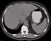
CT image of the liver and abdomen with no contrast enhancement
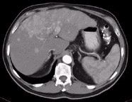
CT image of the liver and abdomen with arterial phase contrast enhancement
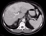
CT image of the liver and abdomen with venous phase contrast enhancement
