


In all original CT scanners (1974 to 1987), the x-ray power was transferred to the x-ray tube using high voltage cables wrapped around an elaborate set of rotating drums and pulleys. The rotating frame (or gantry) would spin 360 degrees in one direction and make an image (or a slice), and then spin 360° back in the other direction to make a second slice. In between each slice, the gantry would come to a complete stop and then reverse directions while the patient table would be moved forward by an increment equal to the slice thickness.
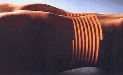
This photo simulates the path that the x-ray beam makes as spiral CT data acquisition of the abdomen is being made. The highlighted area is a man's stomach (man is lying on his back with his arms over his head).
In the mid 1980's, an innovation called the power slip ring was developed so that the elaborate x-ray cable and drum system could be abandoned. The slip ring allows electric power to be transferred from a stationary power source onto the continuously rotating gantry. State of the art CT scanners with slip rings can now rotate continuously and do not have to slow down to start and stop. The innovation of the power slip ring has created a renaissance in CT called spiral or helical scanning.
These spiral CT scanners can now image entire anatomic regions like the lungs in a quick 20 to 30 second breath hold. Instead of acquiring a stack of individual slices which may be misaligned due to slight patient motion or breathing (and lung/abdomen motion) in between each slice acquisition, spiral CT acquires a volume of data with the patient anatomy all in one position. This volume data set can then be computer-reconstructed to provide three-dimensional pictures of complex blood vessels like the renal arteries or aorta. 3D CT images from volume data allow surgeons to visualize complex fractures, for example of facial trauma, in three dimensions and can help them plan reconstructive surgery.
MR, ultrasound and digital x-ray fluoroscopy have all made significant improvements in their ability to image the chest, lungs and abdomen. However, spiral CT has kept computed tomography as the primary digital technique for imaging the chest, lungs, abdomen and bones due to its ability to combine fast data acquisition and high resolution in the same study. CT is also unique in that it can provide detailed information of nearly every organ in the upper abdomen and pelvis in one quick examination.
Spiral CT allows the acquisition of CT data that is perfectly suited to three-dimensional reconstruction. A wide range of software techniques and advanced computer systems are being developed that enable creation of amazing 3D "virtual reality" images.
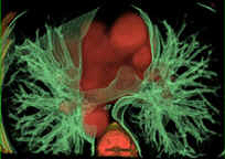
Virtual reality 3-D image of the lungs. The bronchial trees are colored in green and the heart, aorta and vertebrae are colored in red.
