


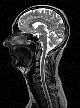
A benefit of MR is that, unlike conventional x-ray or CT imaging, it does not use x-ray radiation. Magnetic resonance imaging is non-invasive and provides exquisite images with excellent contrast detail of soft tissue and anatomic structures like gray and white matter in the brain or small metastatic lesions (cancers) in the liver. In comparison to MR, conventional x-ray provides images of dense structures like bones with good resolution. The x-ray angiogram is the traditional standard for imaging vessels like the carotid arteries in the neck, vessels in the brain, peripheral arm and leg vessels, or the coronary arteries which supply blood to the heart. But conventional angiographic imaging is very labor- and time-intensive and requires administration of significant amounts of contrast to image the blood vessels. X-ray angiography does not provide clear images of the soft tissue organs in the body like the liver or brain (see section on MR Angiography).
Like MR, Computed Tomography (CT) also creates detailed cross sectional images of the body. But, while CT can depict soft tissue structures much better than conventional x-ray, it does not have the contrast detail that MR provides. Many diseases, for example certain brain tumors, are more readily apparent in MR images than in the corresponding CT images due to the better contrast definition of MR.
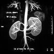
MR imaging is unique in that it can also create detailed images of blood vessels without the use of contrast media (although there is a trend toward the use of special MR-contrast media called Gadolinium when imaging the vessels as well as soft tissue like the brain).
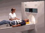
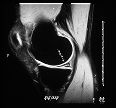
The use of MR imaging as a diagnostic technique continues to grow, allowing the study of more and more body parts. Initially, MR was mainly used to image the brain and spinal column, and each exam could last up to an hour. However, MR scanners can now image a host of additional body parts including injuries of the joints (such as the shoulder, knee, elbow, wrist), the blood vessels (for instance, carotid arteries, renal arteries, peripheral leg arteries), the breast, as well as abdominal and pelvic organs like the liver or male and female reproductive anatomy. MR examinations have also become much faster, in some cases rivaling the speed of spiral CT. Skilled MR operators with the latest equipment can now do complete routine studies (e.g. brain or knee) in as little as ten minutes. In some cases, MR systems equipped with Echo Planar Imaging (EPI) packages can use special emergency or fast protocols to do a basic head study in as little as five to twenty seconds.
