


X-ray imaging is one of the fastest and easiest ways for a physician to view the internal organs and structures of the body. X-ray imaging has been available for 100 years and is an excellent tool for assessing skeletal trauma (e.g. broken bones), for diagnosing the gastro-intestinal system (digestive tract), for high resolution diagnostic imaging of the breasts (mammography), and for comprehensive imaging of the thoracic cavity including the lungs and heart. A host of other applications for x-ray imaging are also available including imaging the kidneys, teeth and jaws, and the fine structures of the ear, nose and throat. X-ray diagnostic imaging still comprises a majority of all diagnostic procedures done on a worldwide annual basis.
Specialized x-ray imaging is the fundamental basis of screening and diagnostic mammography, used to detect and guide treatment of breast cancer. Conventional x-ray imaging still plays an important role in the detection, diagnosis and treatment of heart disease and heart attack.
X-ray imaging is also an important part of bone density measurement for the detection of osteoporosis and also plays a key role in orthopedic surgery and the treatment of sports injuries. X-ray imaging is a mainstay in the detection, diagnosis and treatment of cancer.
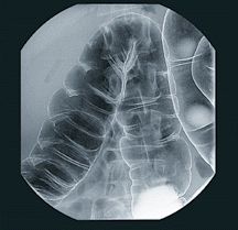
This digital radiograph of the intestine was taken during a GI x-ray examination.
Conventional x-ray imaging encompasses a wide range of techniques and applications. However, in general, x-ray imaging is broken into two major categories:
- Radiographic imaging where a "still image" is made of a bone or organ and shown on film or on a computer screen. A radiograph may be likened to taking a picture with a 35 mm camera.
- Fluoroscopic imaging where a "movie" is made of an organ (for example, swallowing) and viewed on a TV monitor or computer screen.
Several types of radiography and fluoroscopy are available to image the anatomy and function of a wide variety of organs and bones:
- Angiography (imaging of the blood vessels)
- Arthrography (imaging of the joints)
- Barium x-ray (radiograph or fluoroscopy of the gastro-intestinal (GI) tract)
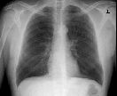
- Chest films (radiograph of the thoracic cavity and heart)
- Cholangiography (imaging of the bile ducts)
- Cholecystography (imaging of the gallbladder)
- Dental X-rays (imaging of the teeth and jaw)
- Lymphangiography (imaging of the lymphatic system)
- Mammography (imaging of the breasts)
- Myelography (imaging of the spinal cord)
- Pyelography (imaging of the urinary tract)
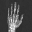
- Skeletal X-rays
- Urography (imaging of the kidneys and bladder)
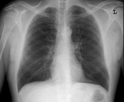
Anterior-posterior (AP) view chest x-ray image
