


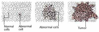
Histology is the study of tissues, including cellular structure and function. Pathologists (physicians who conduct laboratory studies of tissues and cells) often assign a histologic grade to a patient’s cancerous breast tumor to identify the type of tumor present and help determine the patient’s prognosis (projected outcome). The Scarff-Bloom-Richardson system is the most common type of cancer grade system used today. To determine a tumor’s histologic grade, pathologists examine the breast cancer cells and their patterns under a microscope. A sample of breast cells may be taken from a breast biopsy, lumpectomy or mastectomy.
Pathologists closely observe three features when determining a cancer’s grade: the frequency of cell mitosis (rate of cell division), tubule formation (percentage of cancer composed of tubular structures), and nuclear pleomorphism (change in cell size and uniformity). Each of these features is assigned a score ranging from 1 to 3 (1 indicating slower cell growth and 3 indicating faster cell growth). The scores of each of the cells’ features are then added together for a final sum that will range between 3 to 9.
| Tubule Formation (% of Carcinoma Composed of Tubular Structures) | Score |
| > 75% | 1 |
| 10-75% | 2 |
| less than 10% | 3 |
| Nuclear Pleomorphism (Change in Cells) | Score |
| Small, uniform cells | 1 |
| Moderate increase in size and variation 2 | 2 |
| Marked variation | 3 |
| Mitosis Count (Cell Division) | Score |
| Up to 7 | 1 |
| 8 to 14 | 2 |
| 15 or more | 3 |
Courtesy of the American Medical Association
Summary of Histologic Grades of Breast Cancer
A tumor with a final sum of 3, 4, or 5 is considered a Grade 1 tumor (well-differentiated). A sum of 6 or 7 is considered a Grade 2 tumor (moderately-differentiated), and a sum of 8 or 9 is a Grade 3 tumor (poorly-differentiated).
| Grade | Description | Score |
| Grade 1 (lowest) |
Well-differentiated breast cells; cells generally appear normal and are not growing rapidly; cancer arranged in small tubules. |
3,4,5 |
| Grade 2 | Moderately-differentiated breast
cells; have characteristics between Grade 1 and Grade 3 tumors. |
6,7 |
| Grade 3 (highest) |
Poorly differentiated breast cells; Cells do not appear normal and tend to grow and spread more aggressively. |
8,9 |
*Scarff-Bloom-Richardson grade system
Pathologists also look for necrosis (areas of degenerating cancer cells) when determining a tumor’s grade. Cancers with a high grade, necrosis, cancers close to the surrounding margin of breast tissue of a lumpectomy sample, or large areas of DCIS are more likely to recur after breast cancer treatment than other breast cancers.(1)
