


Breast x-rays have been performed for more than 70 years. However, modern mammography has only existed since about 1970, when the first dedicated mammography imaging systems became widely available. Since that time, there has been tremendous advancement in the technology so that today’s examination differs markedly even from those of the early 1980s.
Breast imaging tests, such as ultrasound, sestamibi nuclear medicine, magnetic resonance imaging (MRI), and t-scan (EIS impedance imaging) have been approved by the U.S. Food and Drug Administration (FDA) as supplements to mammography in the detection of breast cancer. Other breast imaging tests such as computerized thermal imaging are currently undergoing clinical trials to determine whether they might be useful in detecting breast cancer, along with mammography.
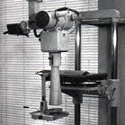 |
| Original dedicated mammography system from 1966 |
- Advances in Mammography
- Digital Mammography
- Computer-Aided Detection System
- Digital Mammography Film Viewer
- Ultrasound
- Magnetic Resonance Imaging (MRI)
- Nuclear Medicine
- T-Scan (EIS Impedance imaging)
- Computerized Thermal Imaging (clinical trials only)
Modern x-ray mammography uses dedicated systems (that is, a machine used only for breast x-rays) to produce x-rays that are high in quality but low in radiation dose. In the past, there were concerns about radiation risks. Modern mammography systems are tightly monitored by the Mammography Quality Standard Act (MQSA) and any risk is far outweighed by the benefits of early breast cancer detection. In order to x-ray the breast, a "softer" type of x-ray is used than for other parts of the body. Mammography is designed for imaging the soft tissue of the breasts as opposed to "harder" x-rays designed to penetrate and image the bones of the body.
X-ray mammography is the only approved examination to screen for breast cancer. However, ultrasound imaging, magnetic resonance imaging (MRI), t-scan (EIS impedance imaging), and nuclear medicine imaging have been further developed in recent years for use in imaging the breast, as adjunct tools to diagnostic x-ray mammography. MRI imaging, ultrasound and nuclear medicine do not provide the spatial resolution (fine detail) available with conventional x-ray mammography. However, MRI provides images with excellent contrast resolution that can help radiologists diagnose and differentiate breast cancers. Ultrasound is useful for imaging cysts and guiding breast biopsy. Nuclear medicine is good for evaluating the spread (metastasis) of cancer into the lymphatic system, other organs and skeletal system.
