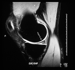
The knee is one of the most complex joints in the body. The rigors of sports require the knee to support motion and tremendous physical stress in a nearly unlimited number of directions. One of the most simple but most important structures of the knee is the cartilage which makes up the smooth bearing-surface of the joint. After years of athletic activity, this cartilage can be slowly worn away. In fact, cartilage loss is one of the most common sports injuries of the knee. Cartilage loss may be caused by gradual thinning and loss of cartilage (osteoarthritis or degenerative joint disease) due to use over time. Traumatic sports injury or similar strenuous activity can cause a meniscal tear (fibrocartilage).

MR image of the knee, side view (sagittal) showing damaged meniscus (arrow)
3D MR Imaging is becoming a powerful tool for assessing cartilage loss in the knee and degenerative joint disease (articular or hyaline cartilage). New orthopedic innovations, such as microfracture are being developed to actually allow regeneration of cartilage in the knee. The process involves pricking tiny holes in the bone near the knee injury. This then causes bone marrow to be released which can spawn the growth of new knee cartilage. Microfracture has been successfully used to rehabilitate the knees of football stars like Bruce Smith, Dan Marino and Rod Woodson. Microfracture also allowed Joe Montana to resume an active, normal life after he limped away from his famed NFL football career. Even weekend warriors have had their active sports lives revived after microsurgery.
Updated: November 2, 2007


