Ultrasound may have a difficult time imaging a breast abnormality that can be felt due to:
- The deep location of the abnormality within the breast
- Operator and equipment factors
- The ultrasound image contrast between the abnormality and the surrounding breast tissue
Ultrasound is not FDA approved as a screening tool for breast cancer for a number of reasons. As stated above, ultrasound cannot reliably detect calcifications, tiny calcium deposits associated with many breast cancers. Ultrasound is also very operator-dependent. That is, the results of an ultrasound exam reflect the ultrasound technologist or the radiologist’s ability to properly manage the equipment. In addition, ultrasound cannot document how much breast tissue has been imaged; therefore, it is difficult to evaluate the thoroughness of the exam. Ultrasound also produces false positive or false negative results from time to time. This means that the ultrasound exam may indicate a breast abnormality when no abnormality is present, or conversely, ultrasound may miss an abnormality. Ultrasound is most valuable when used after mammogram or physical breast exams have indicated an abnormality.
Ultrasound is not a reliable screening tool for breast cancer because:
- It lacks spatial resolution (fine detail)
- It cannot detect most calcium deposits on breast tumors (calcifications)
- Its effectiveness depends largely on the operator
- It cannot document how much breast tissue has been imaged
- False positive or false negative results are possible
Breast ultrasound uses high-frequency waves to image to the breast. High-frequency waves are transmitted from a transducer through the breast. The sound waves echo off the breast; this echo is picked up by the transducer and then translated by a computer into an image that is displayed on a computer monitor.
The ultrasound unit contains a control panel, a display screen, and a tranducer (resembling a microphone or computer mouse). Before the exam begins, the patient will be instructed to lie on a special table. The ultrasound technologist will cover the part of the breast that will be imaged with a gel. The gel will lubricate the skin and help with the transmission of the sound waves.

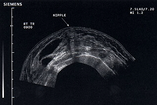
Panoramic ultrasound of the breast showing proliferative breast disease and the lateral predominance of multiple cysts (dark regions on left side of image).
View additional ultrasound images of breast conditions.
When the exam begins, the operator (either the ultrasound technologist or radiologist) will glide the transducer over the breast. The transducer will emit sound waves and pick up the echoes. The computer will then analyze the echoes and display an image on the computer screen. The shape and intensity of the echoes will depend on the density of the breast tissue. If a fluid-filled cyst is being imaged, most of the sound waves will pass through the cyst and emit faint echoes. However, if a solid tumor is being imaged, the sound waves will bounce off the tumor, and the pattern of echoes will be translated by the computer into an image that the radiologist will recognize as indicating a solid mass. Patients may feel a slight pressure from the transducer, but they will not hear the high-frequency sounds.
An ultrasound exam usually lasts between 20 and 30 minutes but may take longer if the operator has a difficult time finding the breast abnormalities being examined. Ultrasound does not use any radiation and is usually pain-free. An ultrasound exam costs much less than a CAT scan or MRI scan.


