

Related Resources
Ultrasound imaging (also called ultrasound scanning or sonography) is a relatively inexpensive, fast and radiation-free imaging modality. Ultrasound is excellent for non-invasively imaging and diagnosing a number of organs and conditions, without x-ray radiation. Modern obstetric medicine (for guiding pregnancy and child birth) relies heavily on ultrasound to provide detailed images of the fetus and uterus. Ultrasound can show fetal development, and bodily function like breathing, urination, and movement. Ultrasound is also extensively used for evaluating the kidneys, liver, pancreas, heart, and blood vessels of the neck and abdomen. Ultrasound can also be used to guide fine needle, tissue biopsy to facilitate sampling cells from an organ for lab testing (for example, to test for cancerous tissue).
Ultrasound imaging and ultrasound angiography are finding a greater role in the detection, diagnosis and treatment of heart disease, heart attack, acute stroke and vascular disease which can lead to stroke. Ultrasound is also being used more and more to image the breasts and to guide biopsy of breast cancer.
Most ultrasound examinations are similar.
- Patient preparation involves removing any articles of clothing or jewelry surrounding the area to be imaged. In some cases, the patient may be asked to wear a patient gown.
- The patient is positioned by the technologist on an examination table. A clear gel (which helps "connect" the ultrasound transducer to the skin) is applied to the area to be examined, for example the abdomen
- The technologist then brings the transducer into contact with the skin and sweeps it back and forth to image the area of interest (e.g the fetal baby). The patient is simply required to relax and stay calm during the examination.
- The technologist will ask the patient to get dressed and wait while the ultrasound images are reviewed, either on film or a TV monitor. In many cases, the technologist or physician reviews the ultrasound images in real time as they are acquired.
- After the ultrasound images are reviewed, the patient will be released from the imaging department or center. In some cases, more images will need to be taken. For more information see "what happens during a diagnostic imaging examination?"
In the 1960's, the principles of sonar (developed extensively by the Defense Department during the second world war) were applied to medical diagnostic imaging. The ultrasound process involves placing a small device called a transducer, against the skin of the patient near the region of interest, for example, against the back to image the kidneys. The ultrasound transducer combines functions like a stereo loudspeaker and a microphone in one device: it can transmit sound and receive sound. This transducer produces a stream of inaudible, high frequency sound waves which penetrate into the body and bounce off the organs inside. The transducer detects sound waves as they bounce off or echo back from the internal structures and contours of the organs. Different tissues reflect these sound waves differently, causing a signature which can be measured and transformed into an image. These waves are received by the ultrasound machine and turned into live pictures with the use of computers and reconstruction software.

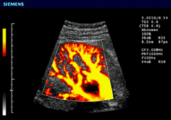
Color ultrasound image of the kidney
Because high-frequency sound waves cannot penetrate bone or air, they are especially useful in imaging soft tissues and fluid filled spaces. Ultrasound is good at non-invasively imaging a number of soft tissue organs without x-rays:
- heart
- pelvis and reproductive organs
- kidneys, liver, pancreas, gall bladder
- eye
- thyroid
- blood vessels
- fetus


