- Prenatal Ultrasound Safety
- Benefits and Limitations of Prenatal Ultrasound
- How Prenatal Ultrasound is Performed
- Types of Transabdominal Prenatal Ultrasound Exams
- Panoramic Prenatal Ultrasound
- Three-Dimensional (3D) Prenatal Ultrasound
- Prenatal Ultrasound Images
- Additional Resources and References
Prenatal ultrasound (also called fetal ultrasound or fetal sonography) has become an almost automatic part of the childbirth process during visits to the obstetrician. It is estimated that up to 70% of women in the United States have prenatal ultrasound exams during pregnancy. Ultrasound is routinely used at 16 to 18 weeks to date the pregnancy and to check the development of the fetus.
The tremendous feelings a mother has for her child growing inside her womb are impossible to describe. The experience is different, yet wonderful, for every mother. The sensation many mothers and fathers feel when they first glimpse live ultrasound images of their fetal babies brings a fascinating reality and a new dimension to the parental experience of pregnancy and childbirth.
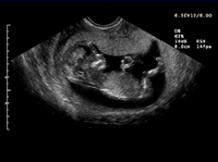
Prenatal ultrasound image showing baby's head, body, arms and legs.
* All prenatal ultrasound images are courtesy of Siemens Medical Solutions.
Prenatal ultrasound has been safely used on pregnant women for over 30 years. The ultrasonic waves used to image the fetus (or other organs) cannot be heard and produce no sensation to the person (or fetus) being imaged. A key benefit of ultrasound is that it does not use x-rays; therefore, it is safe for both the fetus and the mother. The majority of American women have one to three ultrasound examinations during the course of a normal pregnancy.
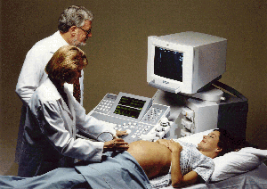
The ultrasound technologist performs the exam while the physician watches the image on the ultrasound monitor.
Modern obstetric medicine (for guiding pregnancy and childbirth) relies heavily on ultrasound to provide detailed images of the fetus and uterus. Ultrasound is an excellent exam for ruling out concerns. However, ultrasound is very operator-dependent. When an experienced physician uses advanced ultrasound equipment, the exam can provide detailed information on the fetus.
However, in some cases, prenatal ultrasound can miss some fetal abnormalities. On average, one third to one half of fetal structural birth defects are not detected with ultrasound. Less commonly, ultrasound can sometimes indicate a fetal abnormality when no abnormality is present, causing stress and worry among the parents. A number of studies have shown that ultrasound is most effective when performed by an experienced physician at a major medical center. When there is an increased risk of genetic or chromosomal birth defects, the physician may order additional testing, such as amniocentesis (sampling of the amniotic fluid around the fetus) or chorionic villus sampling (CVS; sampling the chorionic villi, small tissues that attach the pregnancy sac to the wall of the uterus) in addition to ultrasound imaging.
If ultrasound is needed very early during pregnancy, transvaginal ultrasound, rather than transabdominal ultrasound, is typically performed. Transvaginal ultrasound is performed by inserting a probe into the vagina. Early during pregnancy, the probe is able to get close to the uterus and is helpful in visualizing the fetus. As the pregnancy progresses, transabdominal ultrasound becomes more beneficial than transvaginal ultrasound.
Transabdominal prenatal ultrasound is used to check on the development of the fetus and look for abnormalities. Prenatal ultrasound may be used for the following:
- Determining the age of the pregnancy
- Determining whether the uterus is growing faster or slower than expected and pinpointing the reasons why this might be occurring
- Examining the baby's physical development and functions such as breathing, heartbeat, excretion, and body movement
- Measuring amniotic fluid late in pregnancy
- Imaging the limbs and spinal column to check for proper formation and growth
- Imaging the development of the brain and other major organs
- Determining whether the pregnancy is ectopic (occurring in the fallopian tubes instead of the abdomen)
- Determining whether there is a multiple pregnancy (i.e., twins, triplets, quadruplets)
- Determining the due date of the pregnancy
- Confirming a possible miscarriage (warning signs may include bleeding early in pregnancy or cessation of fetal heartbeat and movement)
- Determining the cause of bleeding during the second or third trimester (causes may include problems with the placenta, which may require a cesarean delivery or special care)
- Diagnosing some birth defects, such as missing limbs, spina defida (a gap in the spine), or malformations of the urinary tract. A special type of ultrasound exam called echocardiography images the blood flow through fetal heart chambers and major blood vessels to help detect heart irregularities.
- Guiding other obstetric/prenatal tests, such as amniocentesis (sampling fluid from the womb) or the sampling of cells from the chorionic villus (vascular projection from the embryo that attaches the pregnancy sac to the uterine wall).
- Guiding prenatal surgery and the safe delivery of medications to the fetus
- Determining whether a cesarean delivery is needed (instances may include when the fetus is especially large or in an abnormal position, or when the placenta is blocking the exit from the uterus)
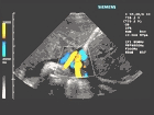
Ultrasound image of umbilical cord
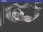
Ultrasound image of intracranial anatomy and facial features
Information obtained from prenatal ultrasound exams can help determine prenatal care, thereby increasing a woman's chances of delivering a healthy baby. Certain problems detected by ultrasound, such as fetal heart rhythms abnormalities or fetal urinary tract blockages, can be treated with medications or surgery before the woman gives birth.
Transabdominal fetal ultrasound is usually a straightforward process that is quick and painless; a typical routine exam (called a basic or level 1 exam) takes approximately 15 to 20 minutes. The woman is usually examined while lying on her back with her belly exposed. First, a jelly-like solution is applied to the skin to help improve the contact between the ultrasound transducer and the skin, allowing clearer images of the fetus. The woman may also be asked to have a full bladder during the ultrasound exam to help with image clarity.
To perform the exam, the ultrasound technologist passes a hand-held transducer across the woman's belly. Inaudible, high frequency sound waves are emitted by the ultrasound transducer and as these sound waves pass into the body, they are reflected back at different strengths and frequencies by the different anatomic structures of both the mother and the fetus. The same transducer receives these reflected sound waves and a computer reconstructs them into real time images.
During the examination, the ultrasound "movie" can often be recorded to videotape or the movie can be frozen (similar to viewing a videotape in "freeze frame" mode) and a still image can be recorded onto paper or film. Typically, moving ultrasound images (images being acquired and displayed at multiple frames per second) give a much better appreciation of the structure of the fetus or other anatomy being imaged than a still image.
Basic or Level I Exam
Routine exam (15 to 20 minutes) to determine pregnancy dates, location of placenta, and check for birth defects.
Targeted or Level II Exam
A longer exam (30 minutes to several hours) that typically uses more advanced ultrasound equipment to further investigate a fetal abnormality. Three
dimensional ultrasound is sometimes used during a targeted exam.
A recent advancement in the field of ultrasound is the ability to scan a panoramic view of the fetus. As the transducer is moved along a part of the body, the image continues to grow, showing the entire scanned area (instead of just showing the area currently being scanned by the transducer). This "panoramic landscape view" gives the obstetrician an appreciation for both fine detail and the "big picture."
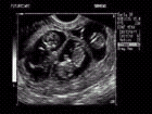
Panoramic ultrasound image of triplet gestation at 11 weeks.
Three-dimensional (3D) ultrasound is another recent advance that can produce 3D images of the fetus that are as detailed as a photograph. This type of imaging may be used during targeted (also called level II) exams when physicians are examining a particular fetal abnormality. Currently, the 3D ultrasound technology is not available on a widespread basis.
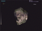
3D ultrasound image showing soft tissue of the fetal face.
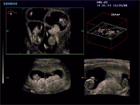
3D ultrasound image of first trimester quadruplets.
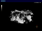
3D ultrasound image of the placenta.
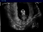
3D ultrasound image of fetal orbits (eye sockets) at 12 weeks.
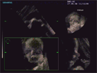
3D ultrasound image showing soft tissue of fetal face.
All images in this Prenatal Ultrasound section are courtesy of Siemens Medical Solutions.
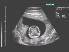
Ultrasound image of 11-12 week fetus shows division of hemispheres and choroid plexus (thin membrane that covers eyeball).
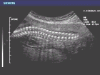
Ultrasound image of fetal spine.
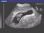
Ultrasound image of first trimester embryo and yolk sac.
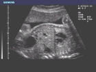
Ultrasound image of fetal liver/lung interface.
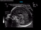
Ultrasound image of intracranial features including the cerebellum and corpus callosum.
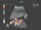
Ultrasound image of blood vessels of umbilical cord.
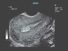
Ultrasound image of vascular changes in the uterus.
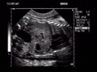
Ultrasound image of fetal bladder, stomach, heart and the liver/lung interface.
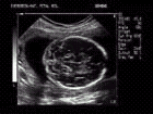
Ultrasound image of fetal intracranial structures including cerebellum.
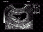
Ultrasound image of yolk sac and fetal pole at 6 week gestation.
- All prenatal ultrasound images are courtesy of Siemens Medical Solutions. Visit Siemens Ultrasound
- The American College of Obstetricians and Gynecologists provides information on prenatal ultrasound and other pregnancy issues. Pamphlets on various aspects of pregnancy, including prenatal ultrasound, may also be ordered through the website.
- The March of Dimes provides information on pregnancy and childbirth, including a detailed section on birth defects.
- General information on ultrasound.
- Information on ultrasound breast imaging.


